

Source: WeChat Official Account “AISRI”
Recently, Peking University Third Hospital performed the world’s first orthopaedic robotic surgery combined with an intraoperative 3D C-arm system for intelligent assisted correction of congenital cervical scoliosis.
For this procedure, confronted with challenges including highly variable anatomy of cervical deformity, adjacent critical neurovascular structures and narrow safe operating space, Tianxuan Xingyao leveraged its intraoperative 3D imaging, intelligent interconnection and instant review capabilities to help the surgical team translate individualized preoperative plans into precise intraoperative manoeuvres, forming an intelligent diagnosis-and-treatment closed loop of “planning — execution — verification”.
Case Background and Clinical Challenges
Patient profile: Hanghang (pseudonym), male, 11 years old. Diagnosis: Congenital cervical scoliosis. Surgical procedure: Robot-assisted correction of congenital cervical scoliosis with combined intraoperative 3D C‑arm system.
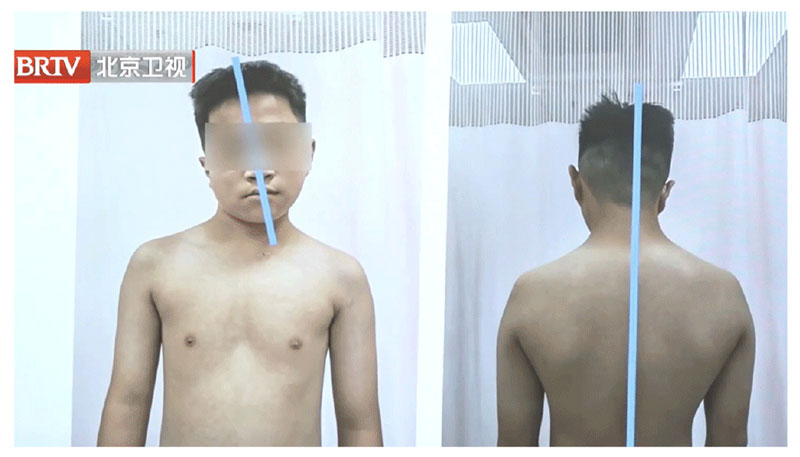
Figure 1 Nation’s First Case! TiRobot & Tianxuan Synergy Empowers Precision Correction for Congenital Cervical Scoliosis
Clinical Challenges
- Complex deformity with marked inter-patient variation: Congenital cervical scoliosis is a rare and complex spinal deformity. Patients show substantial differences in vertebral development, bony fusion, rotational deformity and affected segments, making it hard to rely on personal experience or routine pathways for clinical decision-making.
- High-risk anatomy in the cervical region: The cervical spine sits adjacent to vital structures including the spinal cord, nerve roots and vertebral arteries. The bony corridors of pedicles and vertebral bodies are narrow and may have developmental anomalies. Both internal fixation placement and corrective manoeuvres demand extremely high precision of trajectory and safety margin control.
- The “translation gap” between individualized planning and intraoperative execution: Surgical plans for complex cervical deformities are highly personalized. Translating preoperative precise assessment of deformity anatomy, affected segments and correction strategy reliably and accurately into every intraoperative step is critical to surgical quality.
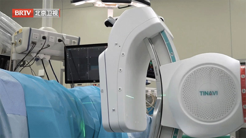
Figure 2 Clinical Challenges, Intelligent Interconnection and Synergy of 3D Image Navigation
PART 01 Intelligent Interconnection: Alignment Between Preoperative Planning and Intraoperative 3D Data
To address the complex bony anatomy and individualized correction requirements of congenital cervical scoliosis, the medical team completed deformity analysis, identification of affected segments and correction trajectory planning before surgery. During the operation, the Tianxuan Xingyao 3D C‑arm interconnects seamlessly with intelligent preoperative planning, intraoperative navigation and the TiRobot surgical robot. It rapidly acquires and transmits 3D image data, providing reliable evidence to convert preoperative plans into intraoperative action.
With 3D spatial information, surgeons can directly identify target segments, vertebrae, pedicles and other key bony landmarks, establishing accurate spatial references for implant placement and corrective procedures.
PART 02 3D Imaging × Navigation × Robot: Visualization of Complex Anatomy to Identify Safety Boundaries
Congenital cervical scoliosis is often accompanied by varying degrees of vertebral developmental anomalies, bony fusion and rotational deformity, which cannot be fully depicted by conventional 2D imaging. Powered by Tianxuan Xingyao’s intraoperative 3D imaging capability, surgeons obtain intuitive 3D volume-rendered images for stereoscopic evaluation of target segments, bony anatomy and critical anatomical relationships. This visualized spatial information delivers clearer imaging evidence for key decisions in managing complex deformities.
Intraoperative 3D images captured by Tianxuan Xingyao serve as fundamental data for the coordinated work of the navigation system and TiRobot. Under navigational guidance, the TiRobot assists surgeons in performing precise operations along planned trajectories, creating a seamless closed loop from “image assessment” to “trajectory planning” and then to “precision execution”.
PART 03 Intraoperative Real-Time Quality Control: 3D Review Provides Documented Evidence for Key Outcomes
In corrective surgery for complex cervical deformity, implant position, insertion trajectory, correction progress and overall spinal alignment require thorough verification. Reliance solely on 2D fluoroscopy or surgeon experience fails to fully demonstrate real outcomes under complex 3D anatomy. At key operative stages and upon completion of correction, Tianxuan Xingyao performs intraoperative 3D review to support the team in verifying the following:
- Position, orientation and spatial distribution of internal fixation implants;
- Relationship between critical bony corridors and surrounding tissues;
- Potential risk signals such as cortical perforation;
- Whether cervical alignment, sequence and overall effect after correction meet expectations.
This closed-loop workflow of “intraoperative acquisition — instant evaluation — adjustment if required — reconfirmation” shifts surgical quality control from postoperative outcome review to key intraoperative milestones, delivering more deterministic safeguards for the treatment of complex cervical deformities. The surgery lasted more than seven hours and was completed successfully. This marks a major breakthrough in the full-process intelligent diagnosis and treatment pathway for congenital cervical scoliosis.
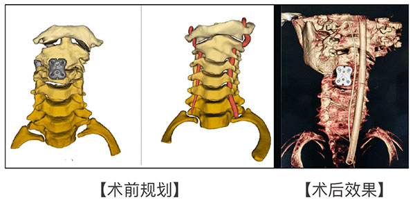
Figure 3 Intraoperative Real-Time Quality Control and Postoperative Outcomes
PART 04 Tianxuan Xingyao Empowers Precision Diagnosis and Treatment of Complex Spinal Deformities
In this world-first procedure, AISRI Tianxuan Xingyao 3D C‑arm, with intraoperative 3D imaging as its core competency, runs through all key stages of complex deformity correction. Evolving from a standalone imaging device into a digital foundational platform for precision surgery, Tianxuan Xingyao reduces information uncertainty in complex operations and improves predictability of clinical decisions and surgical execution through its integrated strengths in “3D imaging, intelligent interconnection and intraoperative quality control”.
Going forward, AISRI Tianxuan Xingyao will continue to leverage high-quality intraoperative 3D imaging to empower spinal, trauma and other complex orthopaedic procedures, offering solid support for the development of precise, intelligent and standardized digital orthopaedics.
Note: All images and videos used in this article are sourced from news reports by Beijing TV.