

Source: WeChat Official Account “AISRI”
Recently, the Tianxuan Xingyao 3D Mobile C‑arm X-ray System, serving as the core device for intraoperative 3D image acquisition, worked in synergy with the TiRobot surgical robot to complete a robot-assisted minimally invasive internal fixation surgery for a right femoral neck fracture. Supported by Tianxuan Xingyao’s 3D imaging capability and intelligent interconnection mechanism, the medical team obtained continuous, quantifiable imaging evidence during key steps including closed reduction and screw insertion. This greatly reduced uncertainty in screw placement and the need for repeated adjustments, bringing the procedure closer to a standardized pathway in terms of efficiency and safety boundary control.
Case Background and Clinical Challenges
Patient profile: Mr. Liu, male, 72 years old. Diagnosis: Right femoral neck fracture. Surgical procedure: Robot-assisted closed reduction and screw internal fixation for femoral neck fracture.
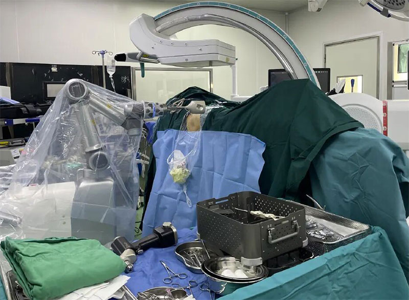
Figure 1 Tianxuan Xingyao 3D Imaging Enables Precise Screw Placement for Femoral Neck Fractures
Clinical Challenges
- High requirements for reduction quality: Femoral neck fractures demand strict alignment, axis restoration and stability. Poor reduction substantially raises risks of internal fixation failure, avascular necrosis of the femoral head and nonunion.
- Low tolerance for placement errors: The femoral neck has a narrow safe corridor; penetration into the joint cavity, extra-cortical perforation and length deviation must be avoided.
- Heavy reliance on conventional fluoroscopy: Closed reduction and screw placement usually require repeated multi-angle fluoroscopy and fine tuning, extending operative time and accumulating radiation exposure for both clinicians and patients.
- Geriatric bone conditions: Elderly patients often have osteoporosis. The holding power of guide wires and screws to bone is highly sensitive to trajectory accuracy, and repeated adjustments may further compromise fixation reliability.
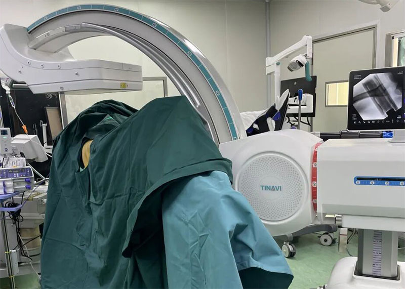
Figure 2 Clinical Challenges, Intelligent Interconnection and Robot Collaboration
PART 01 Intelligent Interconnection: Shortening the Preoperative Preparation Window
In the preoperative planning phase, Tianxuan Xingyao and the Tinavi Sirui orthopaedic surgical robot achieve deep collaboration via Tinavi One Step® for fast imaging data handover. The integrated workflow generates 3D imaging data for planning purposes, to:
- Characterize fracture morphology and the spatial anatomy of the femoral neck;
- Establish spatial references for screw corridors and reduce angle trials that rely purely on 2D fluoroscopy.
Compared with conventional workflows of multiple fluoroscopic positioning and repeated screw trials, the total operative time was reduced by approximately 20 minutes.
PART 02 3D Imaging × Robot Synergy: One-shot Completion of Reduction, Corridor Selection and Screw Placement
Closed Reduction Assessment and Corridor Selection
With high-resolution 3D volume-rendered images from Tianxuan Xingyao, surgeons can directly and stereoscopically evaluate fracture reduction quality and corridor orientation before screw placement, delivering visible evidence for critical clinical decisions.
On this basis, imaging data directly feed into robot trajectory planning and execution. Robot-assisted precise screw placement drastically cuts time cost and operational burden caused by repeated adjustments and improves the success rate of one-time screw insertion, forming a closed loop of “assessment — decision — execution”.
PART 03 Intraoperative Real-Time Quality Control: Closed-loop 3D Review to Improve Controllable Safety Margins
Final Position Review and Risk Screening
After screw insertion, Tianxuan Xingyao performs 3D rechecks to verify key points:
- Whether there is risk of screw penetration into the joint cavity;
- Whether screw position, orientation and overall layout meet stability requirements for fixation;
- Whether warning signs of potential complications such as extra-cortical perforation exist.
This quality-control closed loop of “immediate review — immediate correction” upgrades screw safety assessment from empirical judgment to evidence-based imaging validation.
Against the backdrop of rapid digital and intelligent development in orthopaedics, Tianxuan Xingyao is evolving from a simple imaging device into a digital foundational platform supporting precision surgery. Powered by preoperative and intraoperative intelligent interconnection and data exchange, intraoperative high-resolution 3D imaging and trajectory guidance, plus post-placement real-time quality control and outcome evaluation, the system delivers continuous, quantifiable imaging evidence throughout the whole surgical workflow. It effectively reduces operational uncertainty and improves surgical predictability and reproducibility, offering a practical technical pathway for workflow standardization and risk control in minimally invasive internal fixation for femoral neck fractures.
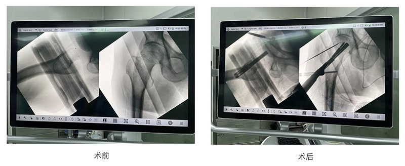
Figure 3 Intraoperative Real-Time Quality Control, Closed-loop 3D Review