

Source: WeChat Official Account “AISRI”
Recently, the Tianxuan Xingyao 3D Mobile C-arm X-ray System assisted the TiRobot surgical robot in completing a highly challenging minimally invasive surgery for an elderly patient with lumbar compression fracture (PVP). During the procedure, leveraging outstanding imaging performance and proprietary intelligent connectivity technology, Tianxuan Xingyao helped surgeons deliver precise treatment down to the millimetre, turning this complex geriatric surgery into a benchmark case of standardized precision medicine.
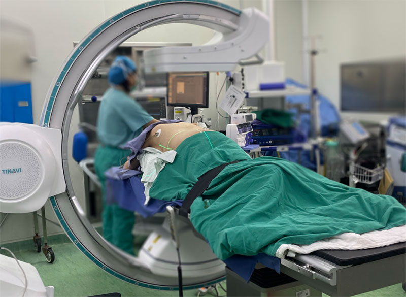
Figure 1 Clinical Perspectives|Tianxuan Xingyao Smart Imaging Advances Minimally Invasive Precision Spine Surgery for Elderly Patients
I. Case Background and Clinical Challenges
Patient profile: Mr. Li, male, aged 80. Diagnosis: Lumbar compression fracture.
Surgical challenges:
- Difficult anatomical identification: Elderly patients often suffer from severe osteoporosis and degenerative changes. Under conventional 2D fluoroscopy, bony structures overlap, blurring boundaries between lesions and adjacent neurovascular tissues.
- Poor surgical tolerance: At age 80, strict limits must be placed on anaesthesia duration and surgical trauma, imposing stringent requirements on operative efficiency.
- High precision demand: Percutaneous puncture must avoid high-risk regions such as the spinal canal; minor deviation may cause irreversible neurological injury.
To address these challenges, the medical team adopted a combined solution of Tianxuan Xingyao 3D Mobile C‑arm + TiRobot surgical robot to successfully perform percutaneous vertebroplasty (PVP). Acting as the “intelligent eye for orthopaedics”, Tianxuan Xingyao resolved clinical hurdles through three key steps.
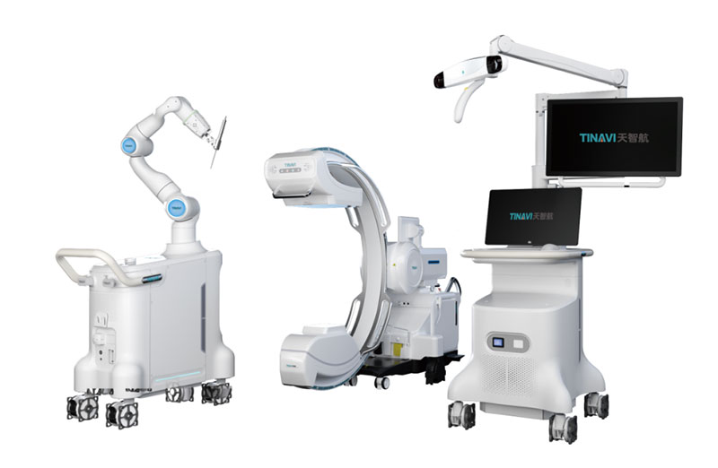
Figure 2 Case Background and Clinical Challenges | Tinavi One Step® Technology
II. PART 01|Tinavi One Step® Technology • Redefining Operative Timing
Preoperative planning: Traditional robotic surgery is often bottlenecked by tedious manual registration, which consumes considerable time and labour. For this case, surgeons activated Tianxuan Xingyao’s exclusive Tinavi One Step® function. No manual calibration phantom is required. Seamlessly interconnected with the robot, the system completes scanning, image reconstruction, registration, data transmission and surgical planning with one click. This integrated workflow saved approximately 30 minutes of planning time and greatly improved surgical efficiency.
III. PART 02|High‑Definition 3D Imaging • Illuminating Blind Spots
Puncture pathway execution: Faced with obscured images caused by osteoporosis, Tianxuan Xingyao rapidly acquires high‑resolution 3D volume rendered images (VRT). It clearly visualises delicate trabecular bone architecture and fracture lines within the vertebral body. Surgeons no longer rely on empirical guesswork. The high-definition 3D imaging delivers real-time reference, enabling surgeons to design an optimal puncture trajectory that bypasses high-risk zones. The puncture needle was placed accurately in a single attempt with no repeated adjustments.
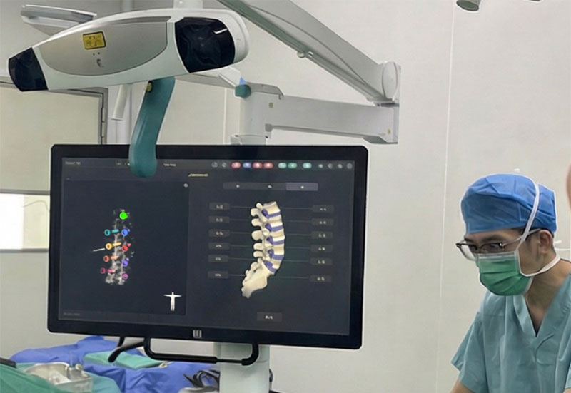
Figure 3 High‑Definition 3D Imaging Illuminates Blind Spots | Intraoperative Real‑Time Quality Control
IV. PART 03|Intraoperative Real‑Time Quality Control • Closing the Efficacy Verification Loop
Cement augmentation confirmation: After bone cement injection, Tianxuan Xingyao immediately performs a 3D scan for review. Images directly demonstrate uniform bone cement dispersion with no intraspinal leakage. The entire workflow runs smoothly and efficiently. Tianxuan Xingyao’s fast, crisp imaging strongly safeguards patient safety while lowering intraoperative radiation exposure.
Closing Remarks: A New Data‑Driven, Imaging‑Led Paradigm for Minimally Invasive Orthopaedic Care
From preoperative intelligent interconnection and data handover, to intraoperative high‑resolution 3D imaging, trajectory guidance, real-time quality control and outcome assessment, Tianxuan Xingyao provides continuous, quantifiable imaging evidence throughout the whole surgical workflow. It effectively reduces operational uncertainty and enhances the predictability and reproducibility of surgery. By advancing standardized management of complex procedures and risk control for high-risk cases, Tianxuan Xingyao helps establish a new data-driven, imaging-led paradigm for minimally invasive orthopaedic diagnosis and treatment.