

Frontline clinicians often share the same question: Why do some C‑arms deliver reliable results no matter who operates them, while others perform inconsistently, depending on the user and sheer luck? On paper, all are 2D C‑arms; yet their real‑world imaging performance can differ dramatically. What truly separates them is not specification sheet figures, but image stability.
What is image stability? Simply put: consistent image quality reliably obtained regardless of the operator, patient or anatomical site. This serves as the core benchmark to judge whether a device is user‑friendly.
Three Most Common Clinical Issues
Issue 1: Image quality varies greatly with different operators. The device seems to “pick its user”. Some operators capture excellent images, while others get overly bright, dark or even non‑diagnostic images.
Issue 2: Inconsistent results for the same patient and anatomical site. Performance becomes a matter of luck. Images may appear sharp one moment, and hazy or dark the next.
Issue 3: Gradual image degradation over time. The device “ages poorly”. Imaging quality looks good at installation but noticeably declines after six months or one year.
These seemingly separate problems stem from one root cause: insufficient system capability.
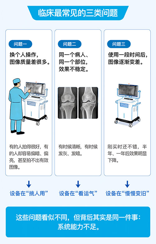
Figure 1 Why do 2D C‑arms vary so greatly in image stability, even among comparable models?
I. Slow Device Response: The “Light Tap” Operation Pattern
The real operational rhythm in operating rooms involves quick foot pedal taps of roughly 0.2–0.5 seconds. Some devices, however, require the pedal to be held for more than two seconds before producing stable images. Consequence: operators who hold the pedal longer get good images, while those tapping quickly receive poor results. In extreme cases, a quick tap triggers no X‑ray output or causes flickering image brightness.
The fundamental cause: the device response speed fails to match clinical workflow. What defines a high‑performance unit? A premium C‑arm should generate consistent high‑quality images even with brief “light taps” as short as 0.2 seconds. This is not a matter of operating habit; it demonstrates inherent system capability.
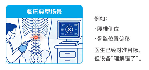
Figure 2 Slow device response and failure to recognize the imaged target
II. The Device “Fails to Understand What You Are Imaging”
Many devices adopt basic brightness control logic, evaluating brightness solely from a small region at the image centre. A typical clinical scenario: minor patient positioning shifts move the target outside the central zone → the system misjudges exposure → automatic exposure control malfunctions → underexposed or overexposed images. For example, in lateral lumbar imaging, the bony anatomy may shift; the surgeon aligns the target correctly, yet the system misinterprets the scene.
How superior systems solve this: advanced platforms automatically identify and track the region of interest, similar to smartphone autofocus. Stable image output is maintained even with positioning offsets.
III. Image Quality Deteriorates With Extended Use
Many hospitals report excellent performance at installation followed by gradual image degradation over time. A common explanation claims “natural device decay”, but this assumption is incorrect. The true cause lies in incomplete control systems lacking closed‑loop regulation. In simple terms: display parameters remain unchanged, yet actual output drifts. For instance, filament aging and minor circuit parameter variations get amplified within the X‑ray system, leading to obvious shifts in image brightness and contrast.
Performance of robust systems: mature units feature comprehensive automatic adjustment functions to compensate for drift, sustaining consistent output long‑term without progressive image degradation.
Contrast Between Standard and Premium Devices
Conventional devices: require prolonged pedal holding; imaging heavily depends on operator technique. High‑performance devices: produce high‑quality images with a quick tap and deliver stable results for all users.
Image stability is ultimately a matter of system capability, not specifications
- Control system capability: ability to deliver stable output within an extremely short timeframe
- Algorithm capability: accurate recognition of the imaged target
- Clinical awareness: genuine alignment with surgeons’ operating habits
Conclusion: What This Means for Hospitals
Selecting a C‑arm with stable image performance brings far more than improved specifications. It delivers higher surgical efficiency, lower learning curves, reduced operator‑to‑operator variability and more predictable imaging outcomes. Great equipment lets surgeons focus on surgery, rather than adapting to the machine.
- prev: Why is surgical time quietly dictated by the 2D C‑arm?2026-10-16
- next: Why are 2D C‑arms used daily, yet rarely fully satisfying?2026-10-16