

Source: WeChat Official Account “AISRI”
There is an interesting observation: the C‑arm brands clinicians remember most are often the ones that draw the most complaints. Truly excellent devices, by contrast, stay “invisible”. They operate like air; you barely notice their presence — until you switch to an awkward unit. Only then do you realize: “Why is today’s procedure moving so slowly?” More often than not, longer operative times stem not from procedural complexity, but from time quietly “stolen” by the 2D C‑arm.

Figure 1 Why is surgical time quietly dictated by the 2D C‑arm?
I. Positioning Time: The Overlooked Hidden Cost
We routinely track time spent placing screws and suturing wounds, yet rarely account for C‑arm positioning. These fragmented seconds add up and define how smoothly the whole surgery proceeds.
- Moments of “getting stuck” When capturing a perfect lateral view for a prone patient, the C‑arm may get blocked as it rotates. Insufficient opening depth and arc depth create problems. For large patients or complex positioning, the C‑arm requires repeated angular adjustments and may even press against sterile drapes — wasting time and raising contamination risks.
Poor alignment between X‑ray tube height and detector ground clearance presents another issue. Even on a C‑arm with generous opening and arc depth, the central X‑ray beam may sit too high above the floor during lateral imaging, or the detector may be mounted too high. This causes the tube to collide with the underside of the operating table or table legs, preventing close access to the patient. The resulting magnified view fails to display enough vertebral levels. Every maneuver to avoid collision consumes precious time.
- A cumbersome handling feel A user‑friendly C‑arm features light mechanical operation with self‑balancing and controlled damping, enabling effortless handling. If technicians must exert full body force to push or pull the unit, or if the gantry drifts downward once the brake is released, operators must constantly steady the arm with one hand while engaging the brake with the other. Fighting against gravity drains extra effort and makes fine adjustments extremely difficult.
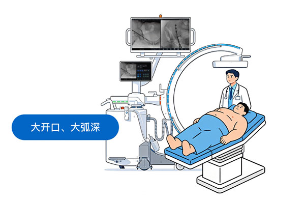
Figure 2 Positioning Time: The Overlooked Hidden Cost
II. The Workflow Killer: Repeated Fluoroscopy and Waiting
Beyond positioning, uncertainty is another major culprit behind lost time.
- The frustration of “We need another shot” The ideal scenario delivers a perfect image with a single exposure. In practice, images often turn too dark or overexposed, requiring parameter tweaks and retakes. C‑arm response lag delays image rendering after foot pedal activation. Surgeons may even doubt whether the pedal registered input, breaking their operative rhythm. Unstable mechanical movement can cause slight positional drift after locking, forcing additional fluoroscopy to reconfirm the target.
- Disrupted clinician-technician coordination Many minimally invasive surgeries require hundreds of fluoroscopic runs or spot exposures. Smooth collaboration between surgeons and technicians is critical. Complicated device controls force technicians to navigate menus repeatedly to access functions, or surgeons must continuously verbalize angle corrections. This communication overhead multiplies total operative duration.
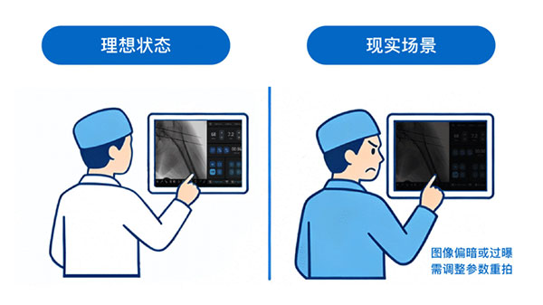
Figure 3 Workflow Killers and Hidden Risks
III. Hidden Concern: Invisible Dose Accumulation
Prolonged surgery directly increases radiation dose. For osteoporotic or obese patients, surgeons may perform dozens of exposures in a seemingly straightforward minimally invasive procedure, just to visualize indistinct pedicles or recheck anatomy after C‑arm drift.
Giving surgeons more options Years ago, a highly respected clinical application expert told us thoughtfully: “In clinical practice, what constitutes a perfect surgical image? You archive 500 images from a case; none have poor quality worthy of complaint, yet none are spectacular enough for marketing use. That is the ideal application. ‘Just enough detail’ defines perfect clinical practice. Every bit of extra image quality comes at the cost of extra dose. An artificially perfect image inevitably demands excessive radiation, which harms both patients and clinicians.”
A truly human‑centered system should offer a broad dose adjustment range. It should include high‑energy modes capable of penetrating dense bone, alongside ultra‑low‑dose and pediatric modes that preserve diagnostic image quality. Unfortunately, many devices rely solely on raising radiation dose in pursuit of perceived “sharpness”.
Conclusion
The best technology is technology you barely notice. A high‑quality 2D C‑arm lets surgeons forget about the equipment and focus entirely on the procedure. When the device becomes “invisible”, surgical time naturally shortens.
- prev: TiMerak Tianxuan • Mobile 3D C‑arm2026-10-17
- next: Why do 2D C‑arms vary so greatly in image stability, even among comparable models?2026-10-16